
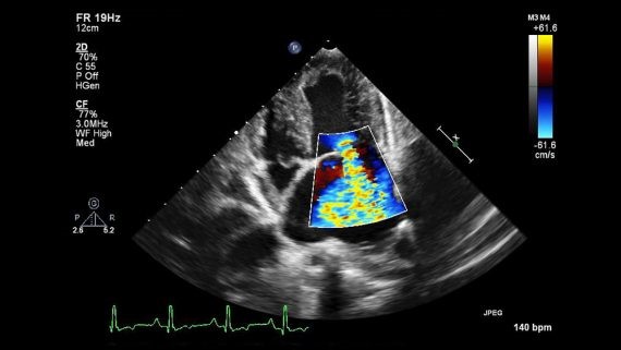
A cardiac triplex (echocardiogram) is a painless and noninvasive test. It uses sound waves (ultrasound) to produce images of the heart. This common test allows the cardiologist performing the test to observe how the heart functions. In this way, it is possible to detect potential heart conditions.
How is a triple heart bypass performed?
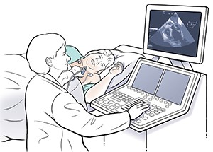
The patient lies on his or her back or on one side, and the cardiologist applies a small amount of special gel to the chest to ensure proper transmission of the ultrasound waves.
The doctor then moves the ultrasound transducer over the skin, capturing real-time images of the heart. These images allow for an assessment of the function of the heart muscle, the valves, and blood flow. During the exam, the patient may be asked to change position or hold their breath for a moment to obtain clearer images.
A cardiac triplex scan usually takes 20 to 30 minutes. After the exam is over, the patient can resume their daily activities immediately.
When should it be done?
A cardiac triplex is recommended for both diagnostic and preventive purposes, depending on the patient’s medical history and symptoms, such as in the following cases:
- Evaluation of valve disorders or disorders of the heart chambers (atria and ventricles)
- Evaluation of potential cardiovascular symptoms, such as shortness of breath or chest pain, which may be of cardiac origin
- Detection of congenital heart defects before birth (fetal echocardiogram)
- Preventive inspection
WARNING:
A standard echocardiogram is one of the most important tests, but it CANNOT detect possible coronary artery disease (CAD). Different tests are needed to investigate it, such as:
- Functional tests (stress test, stress echocardiography, myocardial scintigraphy) for indirect assessment
- Coronary angiography, either invasively (coronary angiography) or using a CT scanner (CT coronary angiography).
However, as mentioned earlier, an echocardiogram performed under conditions of increased cardiac demand—that is, during exercise—can provide important information. Specifically, the stress echocardiogram allows for a reliable assessment of coronary artery function.
What results does a cardiac triplex provide?
A triplex scan allows the cardiologist to accurately assess the structure and function of the heart. The findings of the test are analyzed by the physician. If deemed necessary, they are presented and discussed with the patient as part of the overall cardiological evaluation.
A normal cardiac triplex scan shows no signs of abnormalities in the function of the heart valves, the heart muscle, or blood flow. At the same time, the test may reveal abnormal findings that require immediate treatment or long-term monitoring. For example, it can detect blood clots within the heart chambers and congenital heart defects. In addition, a cardiac triplex scan can reveal diseases of the heart muscle and heart valves, inflammatory conditions of the pericardium, as well as abnormalities of the thoracic aorta.
Types of Echocardiograms
Transthoracic (TTE):
This is the well-known cardiac triplex ultrasound, which is performed in doctors’ offices, diagnostic centers, and hospitals. The technician presses the ultrasound transducer firmly against your skin, directing an ultrasound beam through your chest toward your heart. The transducer records the sound waves reflected from the heart. At the same time, a computer converts the sound waves into moving images that appear on the screen in real time.
Dioesophagus (TOE):
If your doctor wants more detailed images or if it is difficult to obtain satisfactory images with a standard echocardiogram, he or she may recommend a TEE. In this test, a flexible tube containing a transducer is inserted through the mouth (similar to a gastroscopy) and into the esophagus. The transducer is now very close to the heart, with few obstacles in the way, and captures clearer and sharper images. It should be noted that local anesthesia—sedation—is administered to make the examination easier to perform.
Stress Echo (dynamic echocardiography):
will be discussed in a separate chapter, as a stress echo
Does it require preparation?
Preparation for a cardiac triplex depends on the type of test that will be performed. In most cases, no special preparation is required. However, your cardiologist may give you specific instructions, such as avoiding caffeine, alcohol, or smoking for a certain period of time before the test. If you are taking medication, you may be asked to temporarily adjust your dosage of certain medications, but only upon medical advice.
If you are scheduled for a stress echocardiogram, it is recommended that you wear comfortable clothing and shoes. For a transesophageal echocardiogram, fasting and an accompanying person may be required, as the test is performed under sedation. Before the test, the patient is usually asked to remove clothing from the waist up and put on a special medical gown.
Do you need a cardiac checkup?
At the clinics CarE MediC clinics, both cardiac triplex scans and stress echocardiograms on an exercise bike are performed. Cardiac triplex scans are also available during home visits.
Please contact us for more information about the cardiac triplex or to schedule your appointment, either at the clinic or via a home visit.