
WHAT IS IT?
An electrocardiogram (ECG, EKG) is a test that measures the electrical activity of the heartbeat. With each beat, an electrical impulse (or “wave”) “travels” through the heart. This wave causes the heart muscle to contract so that the heart can pump blood. A normal heartbeat on an ECG will show the timing of the atria and ventricles. It is usually one of the first tests performed in a cardiology clinic. In the cardiovascular department of clinics CarE MediC , a 12-lead ECG is performed along with a clinical examination.
WHAT I SEE
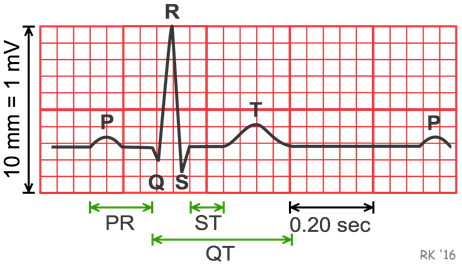
When the electrical impulse is in the right and left atria (upper chambers), the first wave—called the “P wave”—is recorded. This is followed by a short flat line as the electrical impulse travels toward the ventricles. The stimulus then “travels down” to the right and left ventricles, where the next wave—called the QRS complex—is recorded. ” The final wave, or “T wave,” represents electrical recovery or a return to a resting state for the ventricles.
WHY DOES THIS HAPPEN?
An ECG provides important information. First, by measuring the time intervals on the ECG, the doctor can determine how long it takes for the electrical wave to travel through all the chambers of the heart. Measuring the time it takes for a wave to travel from one part of the heart to the next indicates whether the electrical activity is normal, slow, fast, or irregular. Second, by measuring the amount of electrical activity passing through the heart muscle, a cardiologist can determine, for example, whether parts of the heart are abnormally enlarged, or gather other more specialized information. The diagnosis of arrhythmias as well as acute coronary syndromes absolutely requires an ECG recording. It should also be noted that the exercise test is performed with continuous ECG monitoring.
DOES IT HURT?
No.
There is no pain or risk associated with having an electrocardiogram.
IS IT HARMFUL?
No.
The machine only records the ECG. It does not send electricity through the body.